Almost all adults worldwide become seropositive for the virus at some point in time in their lives, but >95% acquire it before 35 years of age. In the case of suspected IM, the heterophile antibody test is used as a screening test. The test is relatively fast, inexpensive, and easy to perform. It, however, is not specific and can be positive for other diseases including acute infections, autoimmune diseases, and cancers.
Also, false negative results are common in children. Further, the heterophile test may be falsely negative in approximately a quarter of patients in the first week of acute illness. EBV-specific antibody tests (antibody testing for viral capsid antigen immunoglobulin M) can be used to confirm the suspected diagnosis of IM in case of a negative heterophile antibody test.
A positive antiviral capsid antigen IgM antibody test is diagnostic of IM. If serologic tests are not helpful (less likely), then molecular analysis by real-time polymerase chain reaction (RT-PCR) and measurement of Epstein-Barr virus viral load can be helpful.
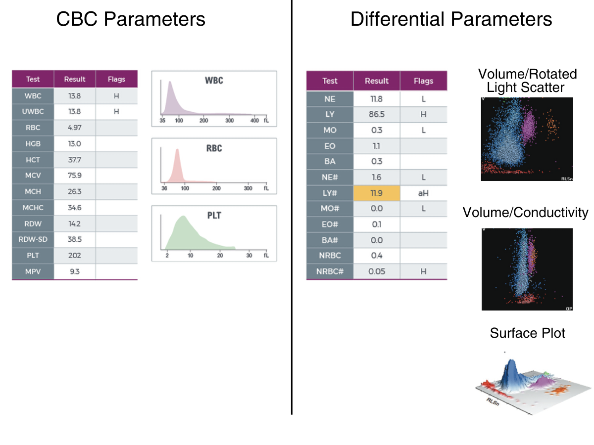
CBC parameters indicate leukocytosis and microcytosis without anemia. WBC histogram shows predominant population in the lymphocyte region. Yellow indicates an action limit is exceeded .Results of the WBC automated differential show neutropenia, monocytopenia, and lymphocytosis. 5PD1 plot (Scatter vs. Volume) and 5PD2 plot (Opacity vs. Volume) show abnormal patterns with predominant lymphocyte population as indicated by instrument messages.
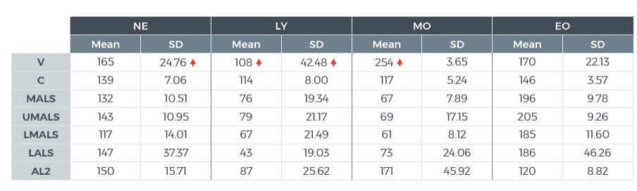
The values of Cell Population Data show increased @LY Volume Mean, increased @LY Volume SD, increased @MO Volume Mean, and increased @NE Volume SD as compared with normal specimens For research use only. Not for use in diagnostic procedures. @ - Research Use Only (RUO) parameters
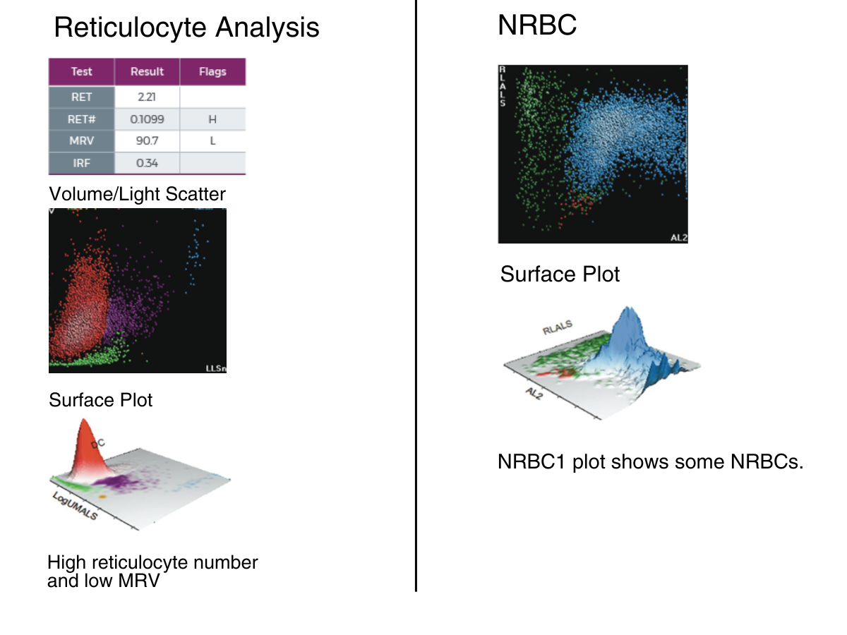
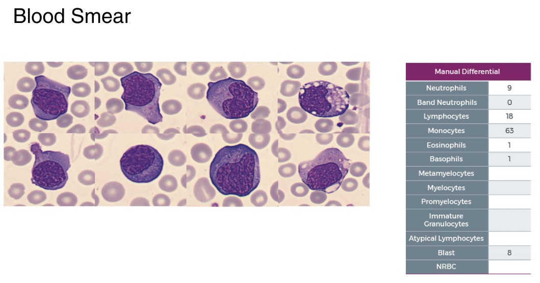
Summary Results
- Microcytosis without anemia, high Retics, and low MRV
- PLT count and PLT histogram are normal
- Leukocytosis with lymphocytosis, neutropenia, and monocytopenia. WBC histogram shows predominant population in the lymphocyte region
- 5PD1 plot (Scatter vs. Volume) and 5PD2 plot (Opacity vs. Volume) show macrocytosis and anisocytosis of lymphocytes, macrocytosis of monocytes, and anisocytosis of neutrophils
- 30.5% of atypical lymphocytes observed on the blood film (83.5% Lymphocytes in total) DIAGNOSIS: INFECTIOUS MONONUCLEOSIS


